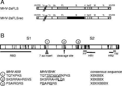
FIG. 1.
Genomic organization of the recombinant viruses. (A) The genome structures of the recombinant MHVs containing either the parental MHV-A59 or the MHV/BHK spike gene and an FL expression cassette are depicted (MHV-2aFLS and MHV-2aFLSrec, respectively). Numbers and lowercase letters designate the genes encoding nonstructural proteins, while genes encoding spike (S) protein, envelope (E) protein, membrane (M) protein, or nucleocapsid (N) protein are marked by the protein abbreviation. The 5′ and 3′ untranslated regions (UTR) are also indicated. (B) The spike protein is depicted as an elongated box. Each vertical line in this box indicates an amino acid substitution in the MHV/BHK S protein compared to the parental MHV-A59 spike protein. The triangle indicates a 7-amino-acid insertion. The MHV-A59 S protein can be cleaved at the position of the arrow into an amino-terminal S1 and a carboxy-terminal S2 domain. Horizontal lines designate the approximate locations of the receptor-binding domain (RBD), putative fusion peptide (FP) (5), heptad repeat region 1 (HR1) and HR2, and the transmembrane domain (TM). The encircled numbers specify the heparin-binding consensus sequences, the locations of which are indicated by gray boxes, while their sequences are given below for the MHV-A59 and the MHV/BHK spike proteins. The amino acid insertions and substitutions in the MHV/BHK spike protein compared to the MHV-A59 spike protein are underlined. The heparin consensus sequences themselves are also shown (X, any amino acid; B, basic amino acid).

