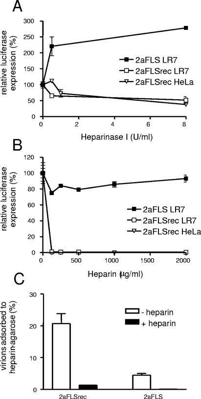
FIG. 4.
Interaction with heparan sulfate/heparin. (A and B) LR7 and HeLa cells were inoculated with the recombinant viruses as described in the legend to Fig. 2A, except that the cells had been pretreated with heparinase I for 1.5 h before the inoculation (A) or the recombinant viruses had been incubated with different concentrations of heparin for 1 h at 4°C (B). At 5 h post infection, the FL activity in the cultures was determined. Standard deviations are indicated. (C) The percentage of MHV virions adsorbed to heparin-agarose beads was determined by a Taqman reverse transcriptase PCR specific for viral genomic RNA, as previously described (11). The black bars (+ heparin) represent the results when virions were incubated with heparin prior to incubation with the heparin-agarose beads, and the white bars indicate the results when the virions were not incubated with heparin prior to incubation (− heparin). Standard deviations are indicated.

