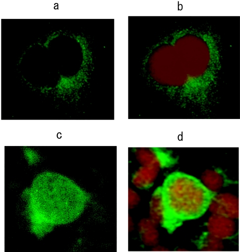
FIG. 5.
Intracellular localization of HBcAg (magnification, ×400). Immunofluorescence staining of HBcAg in HuH-7 cells transfected with (a) pUC19-HBV/E wild-type replicon and (c) pUC19-HBV/E replacement replicon. (b and d) Nuclear staining for the same set of cells was performed by use of DNA staining. DAPI stain shows as red.

