FIG. 1.
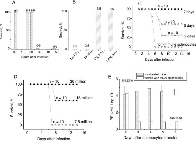
Protective activity of splenocytes from ML29-vaccinated mice. (A) CBA mice were i.p. inoculated with 1,000 PFU of ML29, and at day 7, erythrocyte-free spleen cells were prepared. The recipient mice were i.c. challenged with 1,000 PFU of LASV (time zero), and at different times after challenge, the mice received 30 × 106 immune splenocytes i.v. (B) CBA mice were i.p. inoculated with ML29 at doses varying from 1.5 to 1,500 per animal. At day 7, immune splenocytes were i.v. injected into recipient mice at 3 h after lethal challenge with LASV. (C and D) ML29-immune splenocytes were collected at different time points after vaccination (C) and used at different doses (D) to treat LASV-challenged animals as described above. (E) Tissues from treated and nontreated CBA mice were collected at different time points after challenge and homogenized to prepare 10% (wt/vol) suspensions. Infectious LASV was determined in homogenates by plaque assay. Tissues from four animals were used at each time point. All treated animals survived; nontreated animals died (†) on day 6 after LASV challenge.

