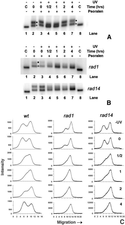
FIG. 4.
(A) Chromatin structure of ribosomal genes during NER in wt cells. Nuclei were isolated from nonirradiated (lane 2) or irradiated (lanes 3 to 7) cells, before (lane 3) and during NER (lanes 4 to 7). After cross-linking with psoralen, DNA was extracted from nuclei, digested with EcoRI, and separated on 1% native agarose gels. As a control (C), DNA was isolated from non-cross-linked nuclei and digested with EcoRI (lanes 1 and 8). After blotting, the filter membranes were hybridized with the random primer-labeled probe (Fig. 1B). Labels at right denote active rDNA (filled circle) and inactive rDNA (open circle). (B) Chromatin structure of ribosomal genes during NER in rad1Δ and rad14Δ strains. Experimental procedures and lanes are as described for panel A. (C) Scan profiles of gels shown in panels A and B.

