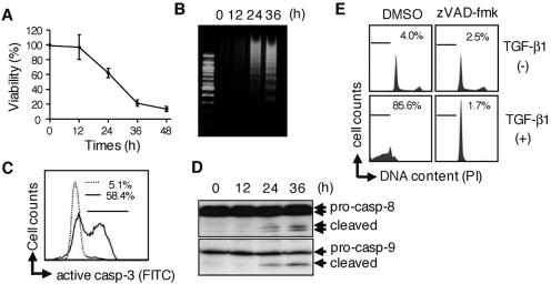
FIG. 1.
TGF-β1 induces caspase-dependent apoptosis in a gastric epithelial cell line, SNU16. (A) Cells were treated with 10 ng/ml TGF-β1 for the period indicated, and cell viability was then determined by quantifying the amount of cellular ATP. The data shown represent the means ± standard deviations (SD) (n = 3). (B) Cleaved genomic DNA was extracted from SNU16 cells treated with TGF-β1 for the period indicated and resolved by 2.0% agarose gel electrophoresis. (C) After incubation with (bold line) or without (dotted line) 10 ng/ml TGF-β1 for 24 h, cells were stained with FITC-conjugated anti-active caspase-3 (casp-3) antibody and activation of caspase-3 was investigated by flow cytometry. Percentages of caspase-3-activated cells are indicated in the figure. (D) After incubation with 10 ng/ml TGF-β1 for the period indicated, cell lysate was analyzed by Western blotting with antibodies to casapse-8 (casp-8) or caspase-9 (casp-9). (E) Cells were pretreated with or pancaspase inhibitor zVAD-fmk (25 μM) for 1 h or left untreated and were subsequently treated with 10 ng/ml TGF-β1 for 48 h. Apoptotic subdiploid cells were analyzed by flow cytometry after staining with PI. Percentages of subdiploid cells are indicated in the figure. (A to E) Data are representative of at least three independent experiments. DMSO, dimethyl sulfoxide.

