
FIG. 3.
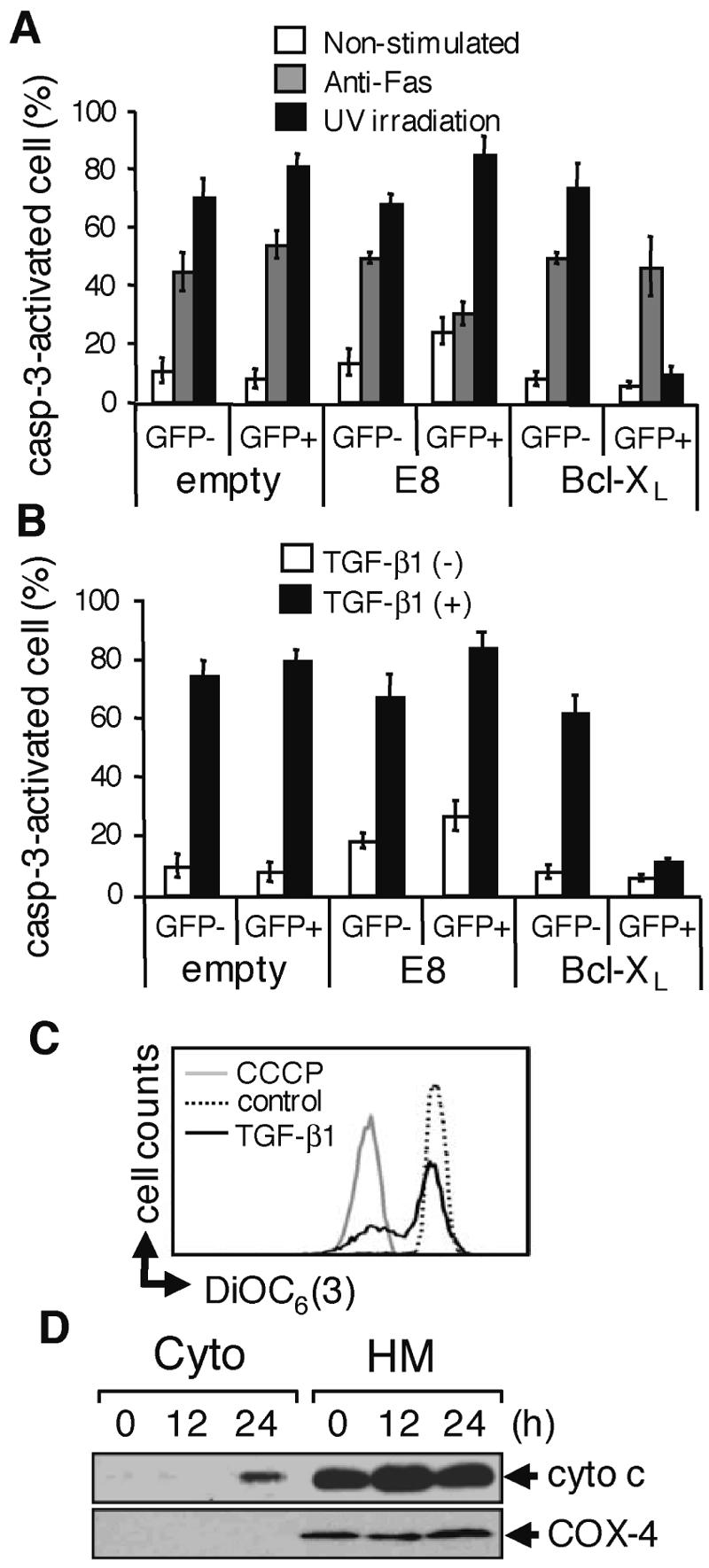
TGF-β1 induces apoptosis in SNU16 cells through the mitochondrial apoptosis pathway. (A) Cells were transfected with the indicated expression vectors (empty or viral FLIP E8 or Bcl-XL; both FLAG tagged) together with a vector for GFP. Transfected cells were stimulated with 1 μg/ml anti-Fas antibody (CH-11) for 12 h or irradiated with 20 kJ/cm2 UV and cultured for 12 h. Caspase-3 (casp-3)-activated cells in the GFP− or GFP+ population were examined as described for Fig. 2D (see Fig. S3B in the supplemental material). Experiments were performed three times, and the means ± SD of caspase-3-activated cells are indicated for the GFP− and GFP+ populations as described for Fig. 2E. (B) Cells transfected as described for panel A were treated with 10 ng/ml TGF-β1 for 24 h and examined as described for panel A (see Fig. S3C in the supplemental material). (C) Cells were treated with 10 ng/ml TGF-β1 for 24 h or left untreated (control) and were stained with DiOC6 (3). As a positive control, cells were similarly analyzed in the presence of carbonyl cyanide m-chlorophenylhydrazone (CCCP). (D) After incubation of SNU16 with TGF-β1 for the period indicated, cytosolic (Cyto) and heavy membrane (HM) fractions were prepared and subjected to Western blotting with anti-cytochrome c (cyto c) antibody. As a control for purity of the subcellular fractionation, the blot was also probed with an antibody to cytochrome oxidase subunit 4 (COX-4).