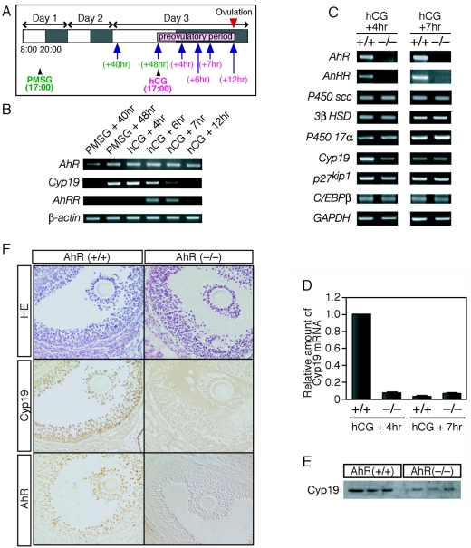
FIG. 4.
AhR regulates the expression of ovarian Cyp19 during the preovulatory period. (A) Schematic representation of the experimental procedure. The estrus cycle was induced artificially by intraperitoneal injection of PMSG at 1700 h on day 1 and of hCG at 1700 h on day 3. Ovaries were collected 40 and 48 h after PMSG injection or 4, 6, 7, and 12 h after hCG injection (indicated by arrows). (B) Profiles of mRNA expression for AhR, AhRR, and Cyp19 during the preovulatory period. Total RNA samples, prepared from ovaries derived from hormone-treated mice at the indicated times (top), were subjected to RT-PCR with primers sets specific for AhR, AhRR, and Cyp19. β-Actin mRNA was used as a control. (C) Expression of mRNAs encoding steroidogenic enzymes and proteins involved in ovarian folliculogenesis. Total RNA samples, prepared from the ovaries of hormone-treated AhR+/+ and AhR−/− mice at the indicated times (top), were used for RT-PCR with the PCR primers. (D) Quantification of Cyp19 mRNA levels. Total RNA samples, prepared from the ovaries isolated 4 and 7 h after hCG injection, were subjected to quantitative RT-PCR analyses. Three animals were used for this experiment. (E) Expression of Cyp19 protein within AhR+/+ and AhR−/− ovaries during the preovulatory period. Whole-cell extracts (10 μg), prepared from the ovaries of hormone-treated (hCG + 5 h) mice, were subjected to Western blot analysis with an anti-Cyp19 antibody. Three AhR+/+ and three AhR−/− animals were used for these experiments. (F) Immunohistochemical staining of Cyp19 and AhR in the granulosa cells of AhR+/+ and AhR−/− ovaries. Five-micrometer paraffin sections were prepared from the ovaries of hormone-treated (hCG + 5 h) mice. Sections were stained with hematoxylin-eosin (HE) or with anti-AhR or anti-Cyp19 antibody.

