FIG. 4.
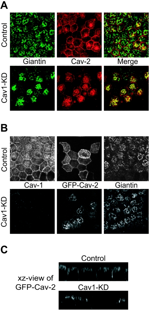
Caveolin-2 is retained in the Golgi apparatus in cav1-KD MDCK cells. (A) Control and cav1-KD MDCK cells were grown on Transwell filters for 3 days. Cells were fixed with paraformaldehyde, permeabilized with saponin and immunostained using giantin and caveolin-2 antibodies to visualize the Golgi apparatus and endogenous caveolin-2, respectively. In control cells caveolin-2 was mainly localized at the basolateral membrane. An intracellular perinuclear staining was also seen. In cav1-KD MDCK cells caveolin-2 accumulated in the Golgi, whereas the plasma membrane pool was strongly reduced. (B) Control and cav1-KD cells were grown on filters for 2 days followed by an infection with an adenovirus expressing GFP-cav2 fusion protein. Sixteen hours later, cells were fixed, permeabilized with saponin, and immunostained using antibodies against caveolin-1 and giantin. In control cells GFP-cav2 was found at the basolateral membrane with some additional perinuclear/subapical intracellular staining. In cav1-KD cells, basolateral staining wa s not observed and GFP-cav2 was retained in the Golgi. (C) xz view of GFP-cav2 in filter-grown control and cav1-KD MDCK cells.

