FIG. 8.
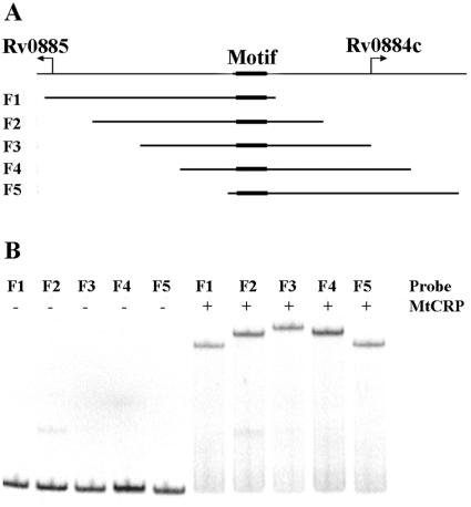
DNA bending by CRPMt. (A) Graphic showing the genetic structure of the Rv0884c-Rv0885 intergenic region. Five 156-bp subregions, designated F1 to F5, were amplified by PCR, with the binding site at a different location within each fragment, as shown. (B) Fragments F1 through F5 were labeled and used for EMSA with 35 nM of CRPMt. Unbound probes showed similar mobilities (left half of gel), while the mobility of each protein-DNA complex varied depending on the position of the CRPMt binding site within the fragment (right side of gel).

