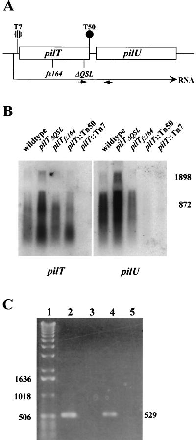
FIG. 8.
Transcription patterns within the pilT-pilU locus in wild-type and mutant strains. (A) Physical map of the locus in the strains analyzed. (B) Northern blot analyses of pilT and pilU expression. Total RNAs (10 μg) from wild-type and mutant strains were electrophoresed on a formaldehyde-agarose gel and blotted onto a nylon membrane. The transferred RNA was hybridized with a pilT probe, yielding the autoradiogram shown on the left. The membrane was then washed and rehybridized with the pilU probe, yielding the autoradiogram shown on the right. The positions of RNA size markers (in base pairs) are shown on the right. (C) Detection of an RNA species spanning pilT and pilU by RT-PCR. Lane 1, 1-kb DNA ladder; lane 2, positive control amplification of intergenic region with wild-type strain chromosomal DNA as the template and with the primers indicated by small arrows in panel A (the expected PCR product is 529 bp long); lane 3, negative control for RT-PCR, in which the conditions were identical to those used to obtain the product in lane 4, except that reverse transcriptase was not added to the reaction mixture; lane 4, RT-PCR with RNA from the wild-type strain; lane 5, RT-PCR with RNA from GT7.

