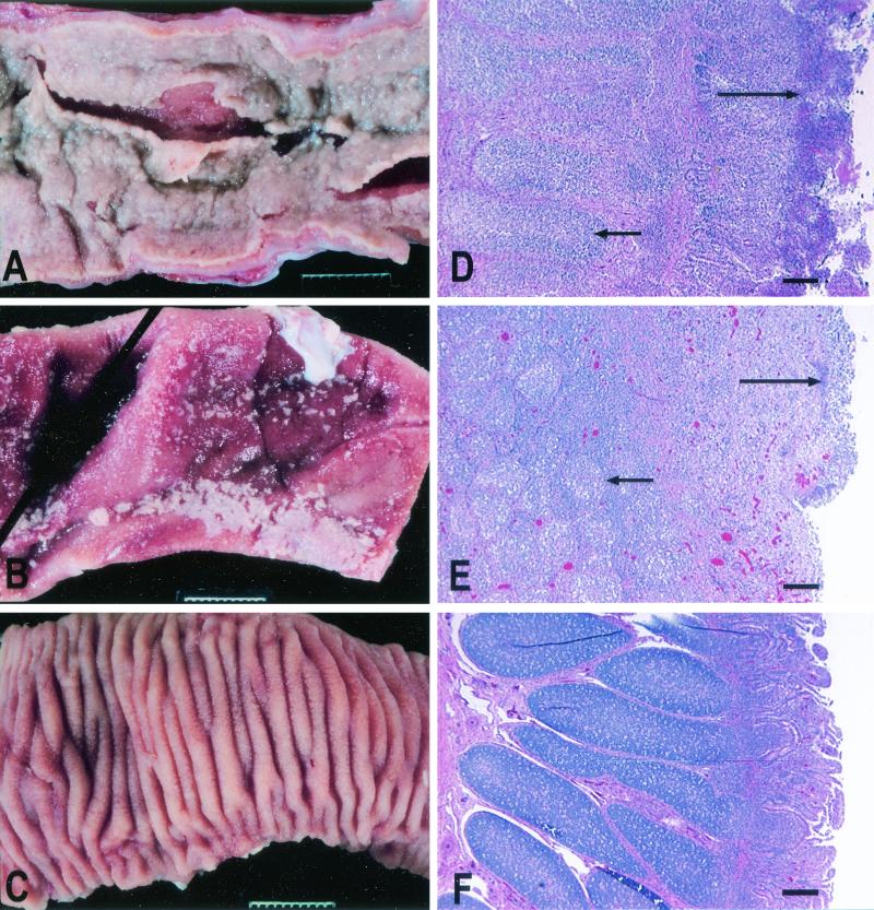
FIG. 8.
Representative examples of the gross pathology and histopathology of Peyer's patches and the terminal ileum of calves inoculated orally with 1010 CFU of different Salmonella serotype Typhimurium strains. (A) Severe acute fibrinopurulent necrotizing enteritis with segmental or continuous pseudomembrane formation in a calf infected with wild-type strain IR715 (similar pathological changes were observed in calves infected with the sopA mutant, the sopD mutant, or the sopE2 mutant). Bar = 1 cm. (B) Marked subacute fibrinopurulent necrotizing enteritis often confined to the Peyer's patches of the terminal ileum of a calf infected with strain ZA10 (sipA). Bar = 1 cm. (C) Normal Peyer's patches and ileum of a calf infected with strain ZA21 (sipAsopABDE). Bar = 1 cm. (D to F) Hematoxylin- and eosin-stained sections of Peyer's patches of calves infected with IR715, ZA10 (sipA), and ZA21 (sipAsopABDE), respectively. The short arrows indicate areas of lymphoid depletion; the long arrows indicate various degrees of fibrinopurulent necrotizing ileitis at the mucosal surface. Bars = 200 μm.

