Abstract
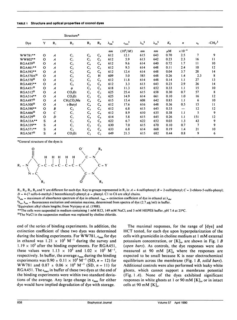
Optical potentiometric indicators have been used to monitor the transmembrane electrical potential (Em) of many cells and organelles. A better understanding of the mechanisms of dye response is needed for the design of dyes with improved responses and for unambiguous interpretation of experimental results. This paper describes the responses to delta Em of 20 impermeant oxonols in human red blood cells. Most of the oxonols interacted with valinomycin, but not with gramicidin. The fluorescence of 15 oxonols decreased with hyperpolarization, consistent with an "on-off" mechanism, whereas five oxonols unexpectedly showed potential-dependent increases in fluorescence at less than 2 microM [dye]. Binding curves were determined for two dyes (WW781, negative response and RGA451, positive response) at 1 mM [K]o (membrane hyperpolarized with gramicidin) and at 90 mM [K]o (delta Em = 0 with gramicidin). Both dyes showed potential-dependent decreases in binding. Changes in the fluorescence of cell suspensions correlated with changes in [dye]bound for WW781, in accordance with the "on-off" mechanism, but not for RGA451. Large positive fluorescence changes (greater than 30%) dependent on Em were observed between 0.1 and 1.0 microM RGA451. A model is suggested in which RGA451 moves between two states of different quantum efficiencies within the membrane.
Full text
PDF
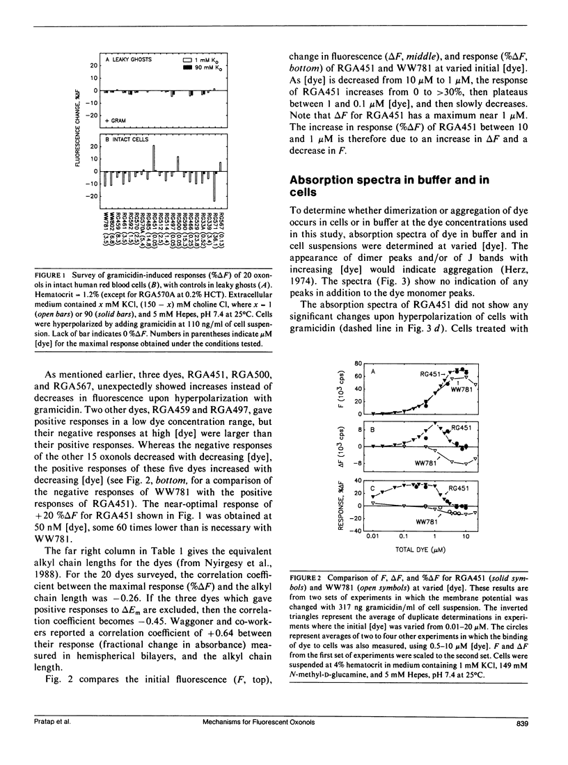
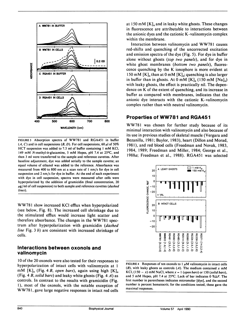
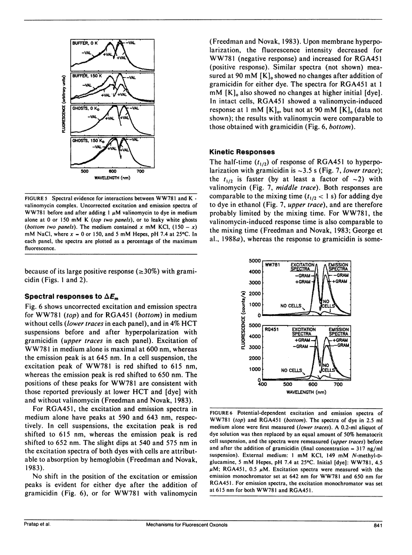
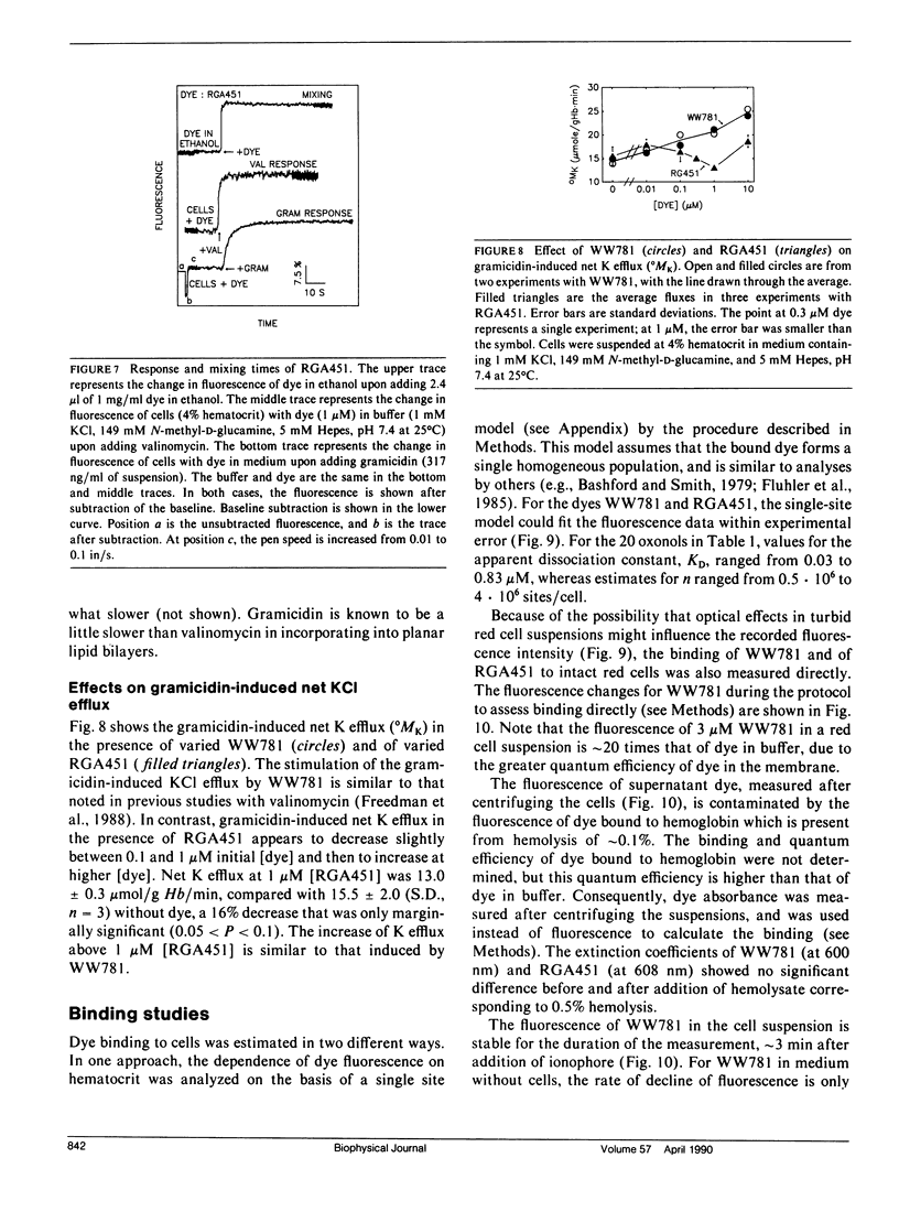
Selected References
These references are in PubMed. This may not be the complete list of references from this article.
- Apell H. J., Bersch B. Oxonol VI as an optical indicator for membrane potentials in lipid vesicles. Biochim Biophys Acta. 1987 Oct 16;903(3):480–494. doi: 10.1016/0005-2736(87)90055-1. [DOI] [PubMed] [Google Scholar]
- Bashford C. L., Chance B., Smith J. C., Yoshida T. The behavior of oxonol dyes in phospholipid dispersions. Biophys J. 1979 Jan;25(1):63–85. doi: 10.1016/S0006-3495(79)85278-9. [DOI] [PMC free article] [PubMed] [Google Scholar]
- Bifano E. M., Novak T. S., Freedman J. C. Relationship between the shape and the membrane potential of human red blood cells. J Membr Biol. 1984;82(1):1–13. doi: 10.1007/BF01870727. [DOI] [PubMed] [Google Scholar]
- Cabrini G., Verkman A. S. Mechanism of interaction of the cyanine dye diS-C3-(5) with renal brush-border vesicles. J Membr Biol. 1986;90(2):163–175. doi: 10.1007/BF01869934. [DOI] [PubMed] [Google Scholar]
- Cabrini G., Verkman A. S. Potential-sensitive response mechanism of diS-C3-(5) in biological membranes. J Membr Biol. 1986;92(2):171–182. doi: 10.1007/BF01870706. [DOI] [PubMed] [Google Scholar]
- DODGE J. T., MITCHELL C., HANAHAN D. J. The preparation and chemical characteristics of hemoglobin-free ghosts of human erythrocytes. Arch Biochem Biophys. 1963 Jan;100:119–130. doi: 10.1016/0003-9861(63)90042-0. [DOI] [PubMed] [Google Scholar]
- Dillon S., Morad M. A new laser scanning system for measuring action potential propagation in the heart. Science. 1981 Oct 23;214(4519):453–456. doi: 10.1126/science.6974891. [DOI] [PubMed] [Google Scholar]
- Dragsten P. R., Webb W. W. Mechanism of the membrane potential sensitivity of the fluorescent membrane probe merocyanine 540. Biochemistry. 1978 Nov 28;17(24):5228–5240. doi: 10.1021/bi00617a024. [DOI] [PubMed] [Google Scholar]
- Fluhler E., Burnham V. G., Loew L. M. Spectra, membrane binding, and potentiometric responses of new charge shift probes. Biochemistry. 1985 Oct 8;24(21):5749–5755. doi: 10.1021/bi00342a010. [DOI] [PubMed] [Google Scholar]
- Freedman J. C., Bifano E. M., Crespo L. M., Pratap P. R., Walenga R., Bailey R. E., Zuk S., Novak T. S. Membrane potential and the cytotoxic Ca cascade of human red blood cells. Soc Gen Physiol Ser. 1988;43:217–231. [PubMed] [Google Scholar]
- Freedman J. C., Hoffman J. F. Ionic and osmotic equilibria of human red blood cells treated with nystatin. J Gen Physiol. 1979 Aug;74(2):157–185. doi: 10.1085/jgp.74.2.157. [DOI] [PMC free article] [PubMed] [Google Scholar]
- Freedman J. C., Hoffman J. F. The relation between dicarbocyanine dye fluorescence and the membrane potential of human red blood cells set at varying Donnan equilibria. J Gen Physiol. 1979 Aug;74(2):187–212. doi: 10.1085/jgp.74.2.187. [DOI] [PMC free article] [PubMed] [Google Scholar]
- Freedman J. C., Laris P. C. Electrophysiology of cells and organelles: studies with optical potentiometric indicators. Int Rev Cytol Suppl. 1981;12:177–246. doi: 10.1016/b978-0-12-364373-5.50015-9. [DOI] [PubMed] [Google Scholar]
- Freedman J. C., Novak T. S. Membrane potentials associated with Ca-induced K conductance in human red blood cells: studies with a fluorescent oxonol dye, WW 781. J Membr Biol. 1983;72(1-2):59–74. doi: 10.1007/BF01870314. [DOI] [PubMed] [Google Scholar]
- Freedman J. C., Novak T. S. Optical measurement of membrane potential in cells, organelles, and vesicles. Methods Enzymol. 1989;172:102–122. doi: 10.1016/s0076-6879(89)72011-5. [DOI] [PubMed] [Google Scholar]
- George E. B., Nyirjesy P., Basson M., Ernst L. A., Pratap P. R., Freedman J. C., Waggoner A. S. Impermeant potential-sensitive oxonol dyes: I. Evidence for an "on-off" mechanism. J Membr Biol. 1988 Aug;103(3):245–253. doi: 10.1007/BF01993984. [DOI] [PubMed] [Google Scholar]
- George E. B., Nyirjesy P., Pratap P. R., Freedman J. C., Waggoner A. S. Impermeant potential-sensitive oxonol dyes: III. The dependence of the absorption signal on membrane potential. J Membr Biol. 1988 Oct;105(1):55–64. doi: 10.1007/BF01871106. [DOI] [PubMed] [Google Scholar]
- Guillet E. G., Kimmich G. A. DiO-C3-(5) and DiS-C3-(5): Interactions with RBC, ghosts and phospholipid vesicles. J Membr Biol. 1981 Mar 15;59(1):1–11. doi: 10.1007/BF01870815. [DOI] [PubMed] [Google Scholar]
- Gupta R. K., Salzberg B. M., Grinvald A., Cohen L. B., Kamino K., Lesher S., Boyle M. B., Waggoner A. S., Wang C. H. Improvements in optical methods for measuring rapid changes in membrane potential. J Membr Biol. 1981 Feb 15;58(2):123–137. doi: 10.1007/BF01870975. [DOI] [PubMed] [Google Scholar]
- Hladky S. B., Rink T. J. Potential difference and the distribution of ions across the human red blood cell membrane; a study of the mechanism by which the fluorescent cation, diS-C3-(5) reports membrane potential. J Physiol. 1976 Dec;263(2):287–319. doi: 10.1113/jphysiol.1976.sp011632. [DOI] [PMC free article] [PubMed] [Google Scholar]
- Hoffman J. F., Laris P. C. Determination of membrane potentials in human and Amphiuma red blood cells by means of fluorescent probe. J Physiol. 1974 Jun;239(3):519–552. doi: 10.1113/jphysiol.1974.sp010581. [DOI] [PMC free article] [PubMed] [Google Scholar]
- Loew L. M., Cohen L. B., Salzberg B. M., Obaid A. L., Bezanilla F. Charge-shift probes of membrane potential. Characterization of aminostyrylpyridinium dyes on the squid giant axon. Biophys J. 1985 Jan;47(1):71–77. doi: 10.1016/S0006-3495(85)83878-9. [DOI] [PMC free article] [PubMed] [Google Scholar]
- Loew L. M., Simpson L. L. Charge-shift probes of membrane potential: a probable electrochromic mechanism for p-aminostyrylpyridinium probes on a hemispherical lipid bilayer. Biophys J. 1981 Jun;34(3):353–365. doi: 10.1016/S0006-3495(81)84854-0. [DOI] [PMC free article] [PubMed] [Google Scholar]
- Nyirjesy P., George E. B., Gupta R. K., Basson M., Pratap P. R., Freedman J. C., Raman K., Waggoner A. S. Impermeant potential-sensitive oxonol dyes: II. The dependence of the absorption signal on the length of alkyl substituents attached to the dye. J Membr Biol. 1988 Oct;105(1):45–53. doi: 10.1007/BF01871105. [DOI] [PubMed] [Google Scholar]
- Ross W. N., Salzberg B. M., Cohen L. B., Davila H. V. A large change in dye absorption during the action potential. Biophys J. 1974 Dec;14(12):983–986. doi: 10.1016/S0006-3495(74)85963-1. [DOI] [PMC free article] [PubMed] [Google Scholar]
- Rothman J. E., Lenard J. Membrane asymmetry. Science. 1977 Feb 25;195(4280):743–753. doi: 10.1126/science.402030. [DOI] [PubMed] [Google Scholar]
- Sims P. J., Waggoner A. S., Wang C. H., Hoffman J. F. Studies on the mechanism by which cyanine dyes measure membrane potential in red blood cells and phosphatidylcholine vesicles. Biochemistry. 1974 Jul 30;13(16):3315–3330. doi: 10.1021/bi00713a022. [DOI] [PubMed] [Google Scholar]
- Tsien R. Y., Hladky S. B. A quantitative resolution of the spectra of a membrane potential indicator, diS-C3-(5), bound to cell components and to red blood cells. J Membr Biol. 1978 Jan 12;38(1-2):73–97. doi: 10.1007/BF01875163. [DOI] [PubMed] [Google Scholar]
- Waggoner A. S. Dye indicators of membrane potential. Annu Rev Biophys Bioeng. 1979;8:47–68. doi: 10.1146/annurev.bb.08.060179.000403. [DOI] [PubMed] [Google Scholar]
- Waggoner A. S., Grinvald A. Mechanisms of rapid optical changes of potential sensitive dyes. Ann N Y Acad Sci. 1977 Dec 30;303:217–241. [PubMed] [Google Scholar]
- Waggoner A. S., Wang C. H., Tolles R. L. Mechanism of potential-dependent light absorption changes of lipid bilayer membranes in the presence of cyanine and oxonol dyes. J Membr Biol. 1977 May 6;33(1-2):109–140. doi: 10.1007/BF01869513. [DOI] [PubMed] [Google Scholar]
- Williamson P., Algarin L., Bateman J., Choe H. R., Schlegel R. A. Phospholipid asymmetry in human erythrocyte ghosts. J Cell Physiol. 1985 May;123(2):209–214. doi: 10.1002/jcp.1041230209. [DOI] [PubMed] [Google Scholar]
- Wolf B. E., Waggoner A. S. Optical studies of the mechanism of membrane potential sensitivity of merocyanine 540. Soc Gen Physiol Ser. 1986;40:101–113. [PubMed] [Google Scholar]


