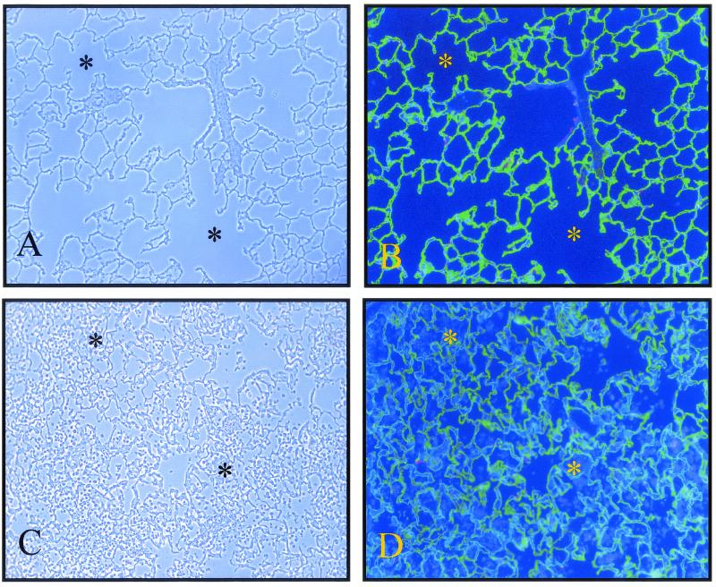
FIG. 3.
Immunofluorescence visualization of leukocyte influx and alveolar epithelium in S. aureus 8325-4-inoculated lungs at 24 h postinfection. Alveolar epithelial type I cells were stained with the anti-RTI40 MAb (green), while alveolar epithelial type II cells were stained with the MMC4 MAb (red). Nuclei were stained with the Hoechst DNA-binding dye. (A) Phase-contrast image of control lung. (B) Corresponding immunofluorescence image. (C) Phase-contrast image of 8325-4-inoculated lung. (D) Corresponding immunofluorescence image. Many of the air spaces (stars) are filled with leukocytes ((Hoechst 33258-only-positive cells) in 8325-4-inoculated lungs in comparison with control lungs. Original magnification, ×100.

