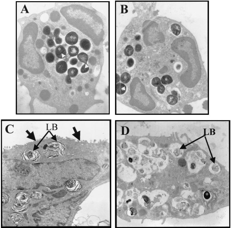
FIG. 8.
Cellular location of S. aureus at the electron microscopic level in 8325-4- and DU5883-inoculated rats. (A and B) Electron micrograph of neutrophils from 8325-4-infected (A) and DU5883-infected (B) lungs. S. aureus (electron dense, approximately 1-μm-diameter particles) cells are internalized into neutrophils from both 8325-4- and DU5883-infected lungs (stars on some internalized S. aureus). The electron-dense line across some S. aureus (arrows in A and B) is a cell wall associated with cell division (37). (C and D) Electron micrograph of an alveolar epithelial type II cell from control (C) and DU5883-inoculated (D) lungs. Type II cell from control lung contains characteristic lamellar bodies (LB) and apical microvilli (arrows) (C). Type II cell from DU5883-infected lung has sloughed from its basement membrane and contains remnants of lamellar bodies (LB) (D). One S. aureus bacterium is located in a vacuole within the cytoplasm of the sloughed type II cell (star) (D). Original magnification: A and B, ×13,000; C and D, ×8,000.

