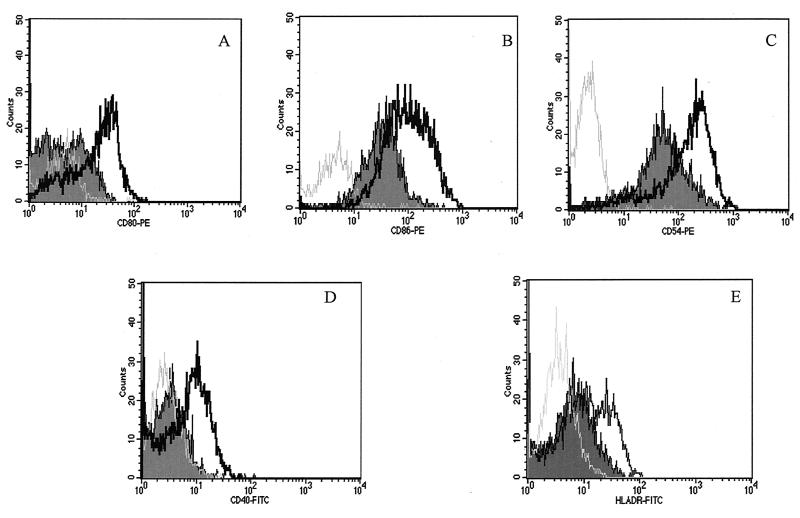
FIG. 3.
Effect of T. cruzi GIPL on costimulatory expression on LPS-stimulated macrophage surfaces. Macrophages obtained from PBMC were incubated in the presence or absence of GIPL (50 μg/ml) plus LPS (500 pg/ml) for 48 h. After this period, the cells were harvested and labeled with specific antibodies to be analyzed by flow cytometry. (A) CD80 expression; (B) CD86 expression; (C) CD54 expression; (D) CD40 expression; (E) HLA-DR expression. The thick black line represents the costimulatory molecule expression from cells stimulated with LPS, the curve that is shaded represents the expression of the costimulatory molecule from cells treated with LPS in the presence of GIPL (Y strain [50 μg/ml]), and the gray line represents cells treated with LPS and stained with isotype antibody controls. The data shown are from a single experiment, representative of five separate experiments.

