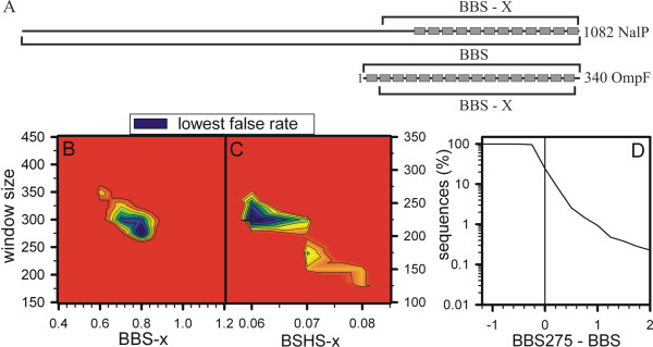
Figure 3.
Analysis of the BBS and BSHS. (A) The β-strand locations of a N. meningitidis (NalP) and an E. coli (OmpF) OM protein are shown. The window for calculating the BBS or a domain based BBS (BBS-x) is indicated. (B-C) The false prediction rate for BBS-x (B) or BSHS-x (C) calculation using different amino acid windows and different cut off scores is shown. The regions with the lowest false prediction rates (black) for the three times weighted pool of the NOM proteins is shown. (D) The percentage of sequences above a certain threshold value of BBS275 minus BBS is shown for the sequences of E. coli.

