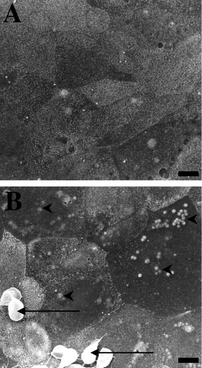
FIG. 1.
Representative scanning electron micrographs of nontransformed human duodenal epithelial SCBN monolayers coincubated with either 5% DMEM growth media (A) or G. lamblia (NF) trophozoites in 5% DMEM (B) for 24 h. Monolayers incubated with G. lamblia trophozoites (arrows) exhibit high incidences of focal surface membrane blebbing (arrowheads) and less distinct microvilli. Similar alterations were observed when monolayers were incubated with NF trophozoite sonicates for 24 h (data not shown). Bars = 5 μm.

