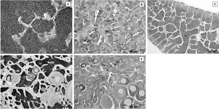
Figure 6.
Pre/postspawning study (adult exposures 1 and 2) showing gonadal histopathology of male and intersex roach. (A and B) Gonads of males with no previous exposure to estrogen. In July (A), the testis was filled with spermatozoa (SZ), and cysts of spermatogonia A (SGA) and spermatogonia B (SGB) were also visible. In September (B), testes of male roach were normal, containing cysts of SGA, SGB, and spermatocytes (SC); there were no obvious differences between the testes of effluent-exposed fish and river water controls. (C–E ) Gonads of males with previous exposure to estrogen. In July, the testis was filled with SZ (C ), and some males were intersex (D). The testis contained SZ, oogonia (O), primary oocytes (PO), and larger oocytes in the cortical alveolus stage (CA). In September (E ), the gonads of these intersex fish contained cysts of SGA, SGB, and SC, together with PO.

