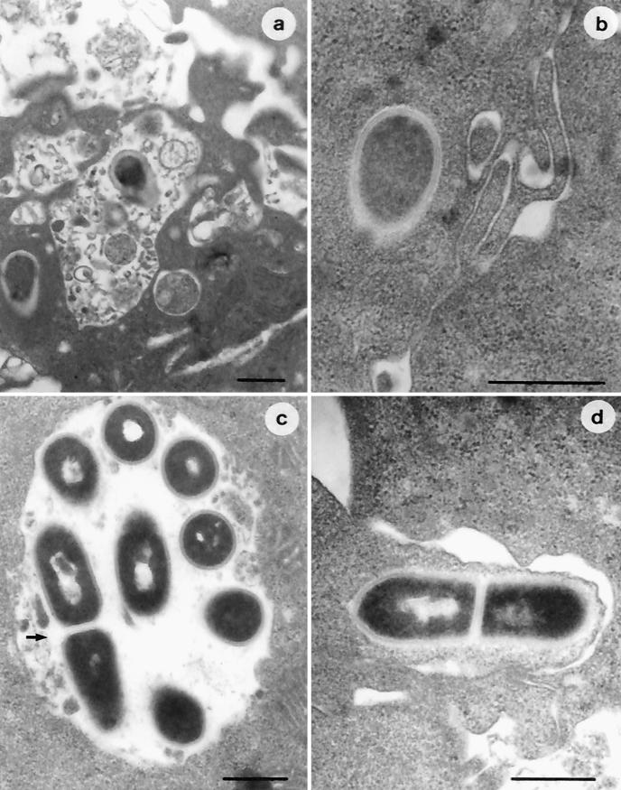
FIG. 5.
Electron micrographs of IFN-γ-activated THP-1 macrophages infected with nonadapted (a and b) and acid-adapted (c and d) L. monocytogenes LM2. A number of nonadapted bacterial cells show dramatic structural damage due to phagosomal digestion (a), whereas only a few cells can be observed free in the cytoplasm (b). Acid-adapted L. monocytogenes cells appear either intact and in active multiplication (arrow) in the phagosome (c) or free in the cytoplasm, with actin tails (d). Bars, 0.5 μm.

