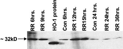
FIG. 7.
Western blot analysis of HO-1 protein in EC during infection with R. rickettsii. EC were left uninfected (Con) or infected for indicated lengths of time (RR), washed thoroughly with PBS, and lysed in cell lysis buffer. A total of 50 μg of sample protein were subjected to polyacrylamide gel electrophoresis. Recombinant HO-1 protein (10 ng) was used as standard. Immunoblotting was performed with a monoclonal antibody reactive with ∼32-kDa HO-1 protein. The standard protein migrates slightly lower on the gel than the native HO-1 protein because of the absence of amino acids 261 to 269 of membrane-spanning region.

