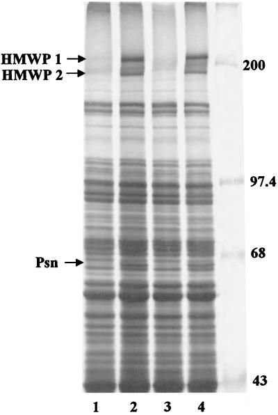
FIG. 6.
SDS-PAGE analysis of whole-cell proteins from Y. pestis strains grown in iron-sufficient and iron-deficient PMH2. Cultures from Y. pestis KIM6+ (lanes 1 and 2), KIM6-2046.1 (irp2::kan2046.1) (lane 3), and KIM6-2086 (irp1-2086) (lane 4) were incubated with 35S-labeled amino acids for 1 h. Total cellular proteins were separated on a 9% polyacrylamide gel and visualized by autoradiography. Cell extracts from iron-deficient cultures (lanes 2 to 4) or iron-sufficient cultures (lane 1) are shown. Sizes of molecular mass markers (in kilodaltons) are indicated. Arrows point to the iron-regulated proteins HMWP1 (240 kDa), HMWP2 (190 kDa), and Psn (68 kDa).

