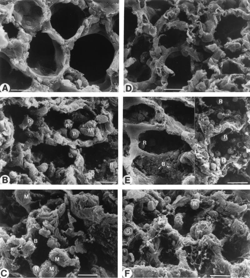
FIG. 10.
Morphological analysis of lung tissues by scanning electron microscopy. Healthy lungs were observed in noninfected immunocompetent (A) and leukopenic (D) mice. Micrographs were taken of lungs from infected immunocompetent mice on days 2 (B) and 3 (C) postinfection and of lungs from infected leukopenic mice on days 2 (E) and 3 (F) postinfection. Abbreviations: B, bacteria; F, fibrosis; M, macrophage; P, polymorphonuclear cell; R, red blood cell. Scale bar = 10 μm.

