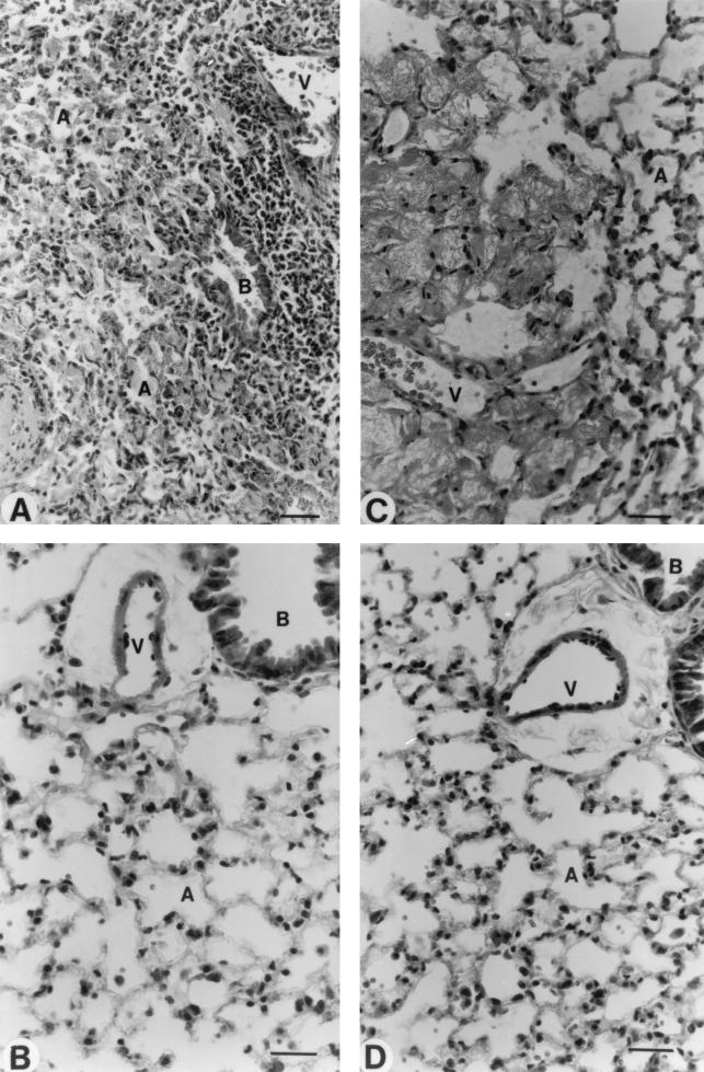
FIG. 9.
Morphological analysis of lung tissues by light microscopy. Studies of infected immunocompetent (A) and leukopenic (C) mice were conducted on day 2 postinfection. Healthy lungs were observed in noninfected immunocompetent (B) and leukopenic (D) mice. Abbreviations: A, alveolar space; B, bronchiolus; V, blood vessel. Scale bar = 20 μm.

