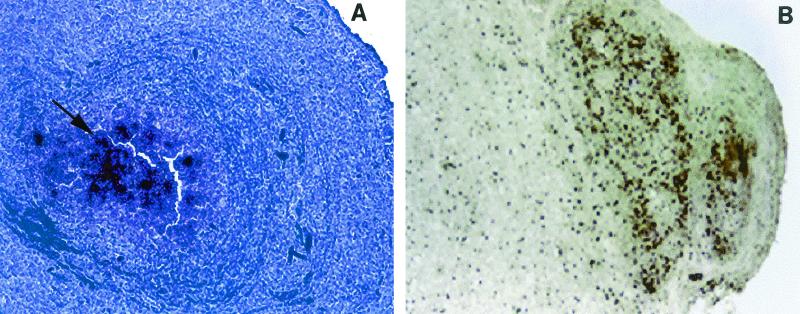
FIG. 2.
Granuloma sections probed by in situ hybridization with 35S-labeled riboprobes for somatostatin. (A) Late-stage granuloma (stage 3), showing numerous positive cells after the section was probed with an antisense probe for somatostatin mRNA. The arrow indicates positive cells overlaid with multiple silver granules. Magnification, ×100. (B) Late-stage granuloma (stage 3), showing numerous positive cells expressing somatostatin protein as revealed by immunohistochemistry. Original magnification, ×200.

