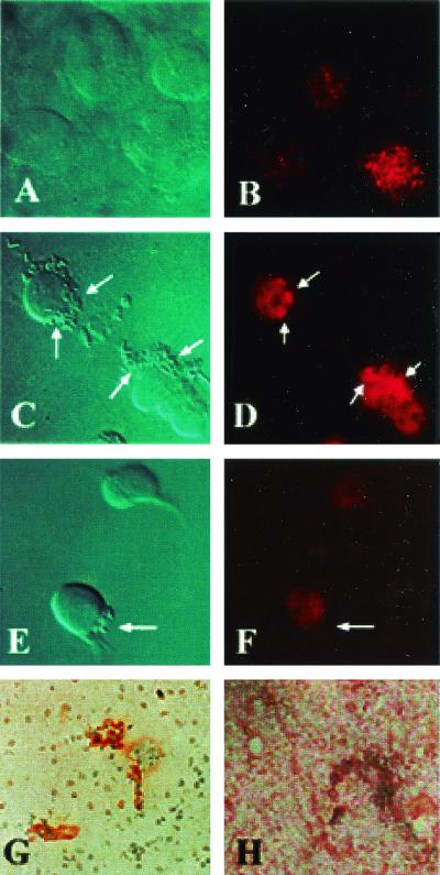
FIG. 7.
Immunocytochemistry of HBMEC and human brain sections. Confluent untreated HBMEC monolayers (A and B) were treated with Ecgp-Ab (1:500), followed by infection with either OmpA+ E. coli (C and D) or OmpA− E. coli (E and F). The monolayers were washed and fixed with 2% paraformaldehyde. The bound Ecgp-Ab was identified by Cy3-conjugated secondary antibody, followed by visualization with laser confocal microscope. The sections of brain embedded in paraffin were stained with either Ecgp-Ab (G) or control rabbit IgG (H), followed by the addition of horseradish peroxidase-conjugated secondary antibody. The brown color was developed with diaminobenzidene and hydrogen peroxide. The pictures were edited and labeled using Adobe Photoshop 6.0. The arrows indicate either the location of invading bacteria or the clusterization of Ecgp.

