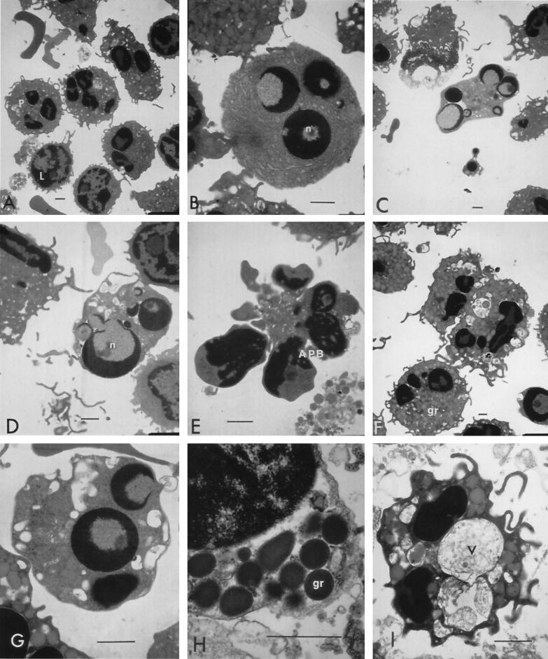
FIG. 5.
TEM of bovine peripheral leukocytes treated with low concentrations of leukotoxin. Normal PMNs (P) and lymphocytes (L) appeared among untreated cells (A). Chromatin condensation was noticed in single or multiple nuclei (n), marginated as rounded or crescent-shaped structures in monocytes (B) and neutrophils (C and D). Apoptotic bodies (APB) were found with their characteristic condensed nuclei surrounded by a thin cytoplasm (E). Cytoplasm of phagocytes showed peripheral translocation of granules (gr; H) and vacuolation (F and G), with vacuoles (v) sometimes appearing large and containing material similar to the extracellular contents (I). (A) Untreated cells; (B to C and F to H) cells treated with 0.2 U of leukotoxin/ml; (D) cells treated with 0.02 U/ml; (E and I) cells treated with 20 U/ml. Bars (all panels), 1 μm.

