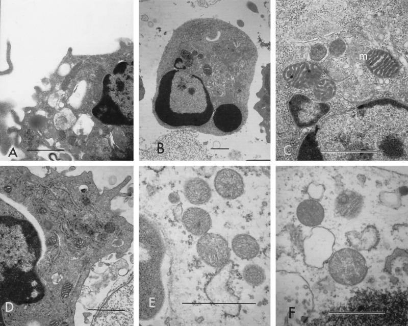
FIG. 6.
Cytoplasmic organelles from cells undergoing apoptosis versus cells undergoing necrosis. Mitochondria (m) and endoplasmic reticulums (er) appeared as condensed, electron-dense structures with discernible internal architecture (A to D), whereas mitochondria in cells undergoing necrosis appeared as light-staining, swollen structures with minimal internal structures (E and F). (A to C) Cells treated with 2 U of leukotoxin/ml; (D) cells treated with 20 U/ml; (E) cells treated with 625 U/ml; (F) cells treated with 2,000 U/ml. Bars (all panels), 1 μm.

