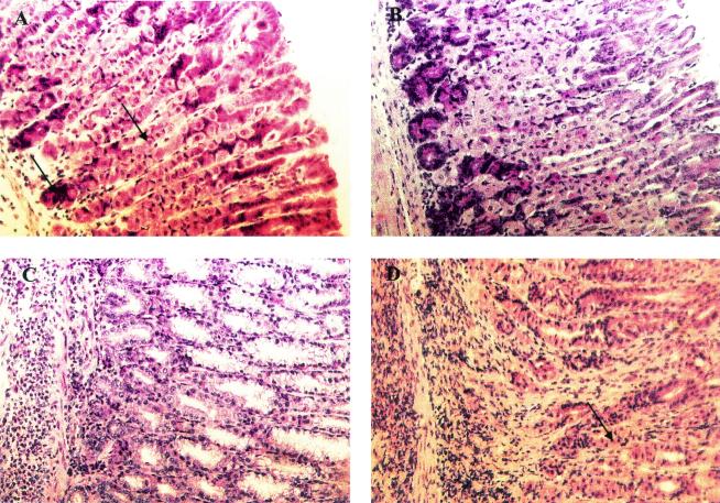
FIG. 3.
Histopathology of mice infected with H. pylori. (A) Oxyntic mucosa of an infected mouse (6 to 8 weeks old) displaying normal morphology. Chief cells and parietal cells in the oxyntic mucosa can be seen (arrows). (B) Mucosa of a mouse treated with CT and examined after reinfection challenge, showing a normal morphology comparable to that of infected mice except for mild focal inflammation. (C) Mucosa of an infected mouse vaccinated with lysate plus CT showing massive lymphocyte infiltration and destruction of chief cells and parietal cells, which are replaced by undifferentiated mucus-secreting cells. (D) Mice immunized with lysate plus CT after eradication and reinfection showing severe inflammation but also the reappearance of some parietal cells (arrow).

