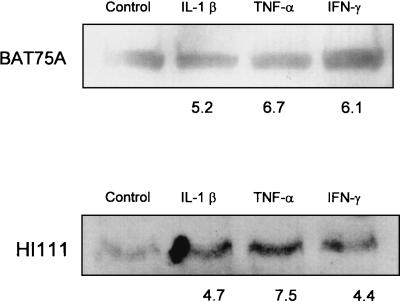
FIG. 3.
Western blot analysis of LFA-1 expression by bovine PMNs incubated with inflammatory cytokines. Freshly isolated bovine PMNs (1 × 10⁶ cells/ml) were incubated with recombinant bovine IL-1β (50 ng), recombinant human TNF-α (50 ng), recombinant bovine IFN-γ (50 ng), or medium (control) for 60 min at 37°C. Total cell lysates were prepared, and equal amounts of total protein were loaded onto Tris-HCl-4 to 20% polyacrylamide gradient gels, electrophoresed, and transferred to nitrocellulose membranes. The membranes were blocked, washed, and probed with anti-LFA-1 MAb BAT75A or HI111 at 37°C for 1 h. The blots were washed and probed with horseradish peroxidase-conjugated anti-mouse IgG at 37°C for 1 h. Immunoreactive proteins were visualized with a SuperSignal West Pico chemiluminescence kit, and relative band intensities were determined by using ImageQuaNT software. The numbers below the bands are the mean fold increases in LFA-1 expression by bovine PMNs incubated with the cytokines compared with the expression by unstimulated PMNs (three separate experiments).

