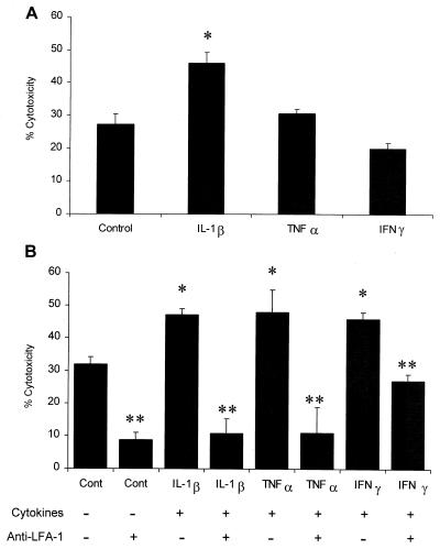
FIG. 5.
Incubation of bovine PMNs with inflammatory cytokines enhances LKT cytotoxicity. Freshly isolated bovine PMNs (1 × 10⁶ cells/ml) were incubated with recombinant bovine IL-1β (50 ng), recombinant human TNF-α (50 ng), recombinant bovine IFN-γ (50 ng), or medium (control) for 15 min (A) or 1 h (B) at 37°C. In the latter experiments (B) some of the cells were incubated with an anti-LFA-1 MAb (BAT75A) (final concentration, 50 μg/ml) for 40 min before addition of LKT. Control and treated PMNs were then plated in 96-well plates and incubated with partially purified M. haemolytica LKT (1 U) for 1 h at 37°C. Cell viability was assessed by XTT reduction. The data are the means ± standard errors of the means for four independent experiments. One asterisk indicates that the value for cytokine-stimulated cells was statistically significantly greater than the value for control cells (P < 0.05). Two asterisks indicate that the value for anti-LFA-treated cytokine-stimulated PMNs was significantly less than the value for untreated cytokine-stimulated PMNs.

