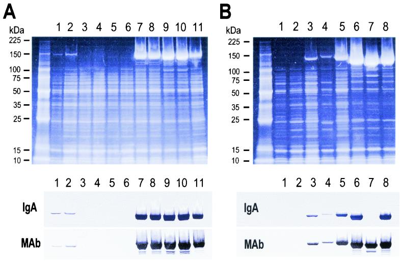
FIG. 1.
SDS-PAGE profiles of cell-bound proteins of GBS of RDP Ia-3 and Ib-1. SDS-solubilized proteins from strain no. 5 to 15 (lanes 1 to 11) of RDP Ia-3 (A) and strain no. 16 to 23 (lanes 1 to 8) of RDP Ib-1 (B) were analyzed with 5 to 20% gradient gels. For each RDP, SYPRO Red staining and immunoblotting are shown. The proteins separated were blotted onto a polyvinylidene difluoride membrane and probed with human IgA (IgA) or with an anti-β monoclonal antibody (MAb). Numbers at left indicate molecular mass standards.

