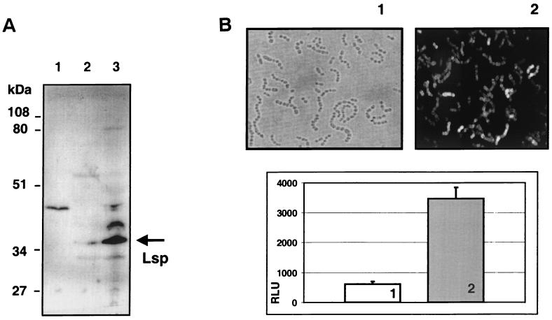
FIG. 2.
(A) Western immunoblot of cell fractions with a polyclonal anti-Lsp antiserum and an anti-rabbit IgG-alkaline phosphatase conjugate. In lanes 1, 2, and 3, 30 μg of total protein from culture supernatant, cytoplasm, and the bacterial envelope fraction, respectively, were separated by SDS-PAGE, transferred to PVDF nylon membranes, and reacted with rabbit anti-Lsp antiserum. The Lsp protein band predominantly visible the cell envelope fraction is marked by an arrow. Relative molecular masses of the marker proteins are shown at the side of the blot. The minor 45-kDa bands in the culture supernatant and cell envelope fractions probably result from a surface-expressed and protease-released IgG Fc-binding GAS protein. (B) Direct immunofluorescence detection of surface expressed Lsp protein in whole GAS bacteria. Rabbit serum obtained before (panel 1) and after (panel 2) immunization with recombinant Lsp protein was for used for direct immunofluorescence detection of Lsp employing qualitative immunofluorescence microscopy (upper two panels) and quantitative measurement in a Cytofluor fluorescence reader (lower panel). RLU, relative light units.

