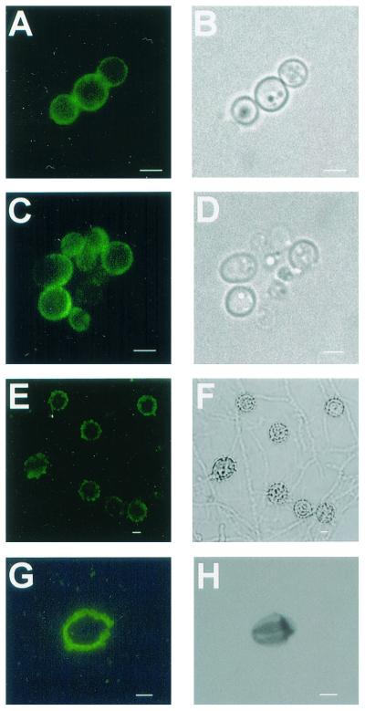
FIG. 4.
Corresponding immunofluorescence (A) and brightfield (B) images of pigmented H. capsulatum strain CIB 1980 yeast cells and the particles isolated from the cells following treatment with enzymes and chemicals (C and D). Corresponding images of mycelial growth of H. capsulatum strain 17262 (E and F) and particles isolated from pigmented conidia (G and H) are shown. Bars, 1 μm.

