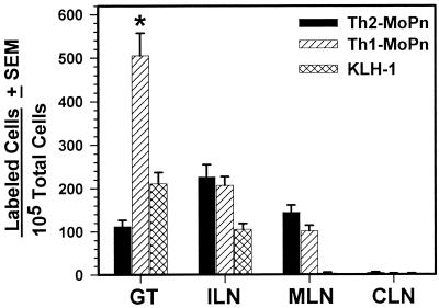
FIG. 3.
Abilities of Th1-MoPn and Th2-MoPn to migrate to the GT 7 days after MoPn infection. Each clone was labeled with PKH-26, and 5 × 10⁶ cells were intravenously transferred into mice infected for 7 days with MoPn. Separate mice received unlabeled cells as a control. Single-cell suspensions were isolated from the tissues 18 h after transfer and analyzed by flow cytometry. Negligible numbers of labeled positive cells (0 to 4 cells) were detected in control mice, and these values were subtracted from those obtained for mice which received labeled cells. The values are means ± standard deviations for two separate experiments in which five mice per group were used for each experiment. An asterisk indicates that a value is significantly greater than the value obtained for clone Th2-MoPn (P < 0.0001). CLN, cervical lymph nodes.

