
Fig. 1.
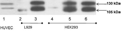
Overexpression of human T-cadherin in HEK 293 and L929 cell lines and constitutive expression of T-cadherin in HUVEC. Whole cell lysates (50 μg/lane) from T-cad-transfected L929 cells (clone TC3-lane 3) and HEK293 cells (clone T5-lane 5, clone T8-lane 6) and mock transfected cells HEK293 (clone HEK/GFP-lane 4) and L929 (clone K9-lane 2) were analyzed using immunoblot method. Lysates of HUVEC, which contain both 105 kDa and 130 kDa forms of T-cadherin (lane 1) served as a positive control.