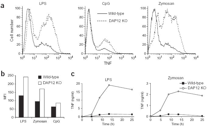
Figure 2.

DAP12-deficient macrophages produce more TNF after TLR stimulation. (a,b) Bone marrow–derived macrophages from wild-type or DAP12-deficient (DAP12 KO) mice were incubated with LPS (0.4 ng/ml), CpG DNA (0.04 μM) or zymosan (ten particles per macrophage) for 4 h (CpG DNA and zymosan) or 14 h (LPS) in the presence of Brefeldin A for the final 4 h. TNF secretion was then assessed; data are represented by one-parameter histograms (a) or by the mean fluorescent intensity (MFI) of the TNF-producing population (b). Data are representative of four independent experiments. (c) Bone marrow–derived macrophages (day 7) were stimulated with LPS (0.5 ng/ml) or zymosan (ten particles per macrophage). Supernatants were removed at the indicated time points, and TNF concentrations were measured using ELISA. Data are representative of three independent experiments.
