
Figure 5.
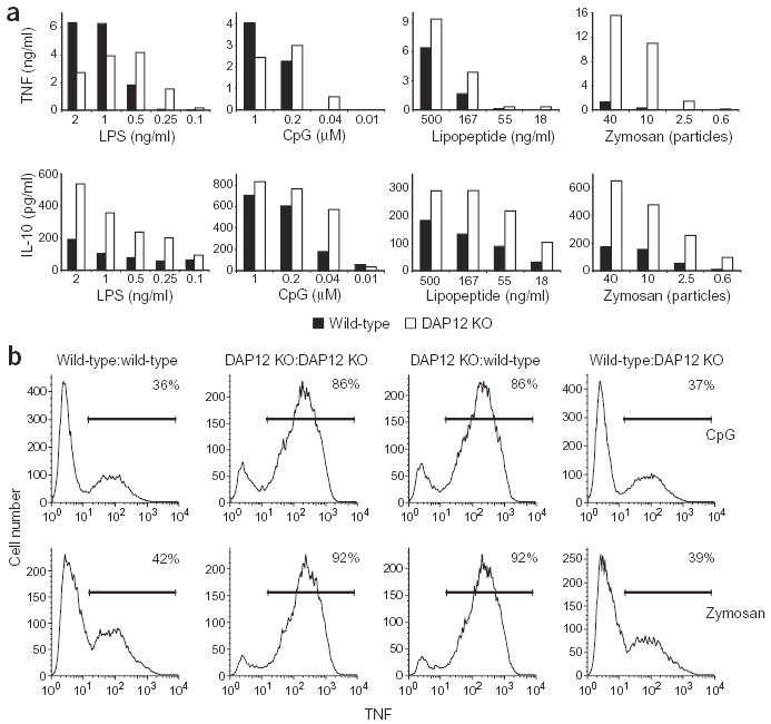
Soluble inhibitory factor production does not explain differences between wild-type and DAP12-deficient macrophages. (a) Bone marrow-derived macrophages from wild-type and DAP12-deficient (DAP12 KO) mice were stimulated with the indicated concentrations of LPS, zymosan, CpG DNA and lipopeptide for 16 h. Supernatants were then assayed for TNF or IL-10 using ELISA. (b) Wild-type and DAP12-deficient macrophages were cultured in the top and bottom chambers of culture plates separated by a porous membrane. Cells in both top and bottom chambers were treated with CpG DNA (0.01 μM) or zymosan (ten particles per macrophage) for 4 h, and TNF-producing cells in the bottom chambers were determined by using flow cytometry (Fig. 2a). In the histograms shown, the type of cells in the top and bottom chambers of the culture plates are indicated as cells in bottom chamber:cells in top chamber. The percentage of TNF-positive cells is indicated in the top right corner of each histogram. Data shown are representative of two independent experiments.