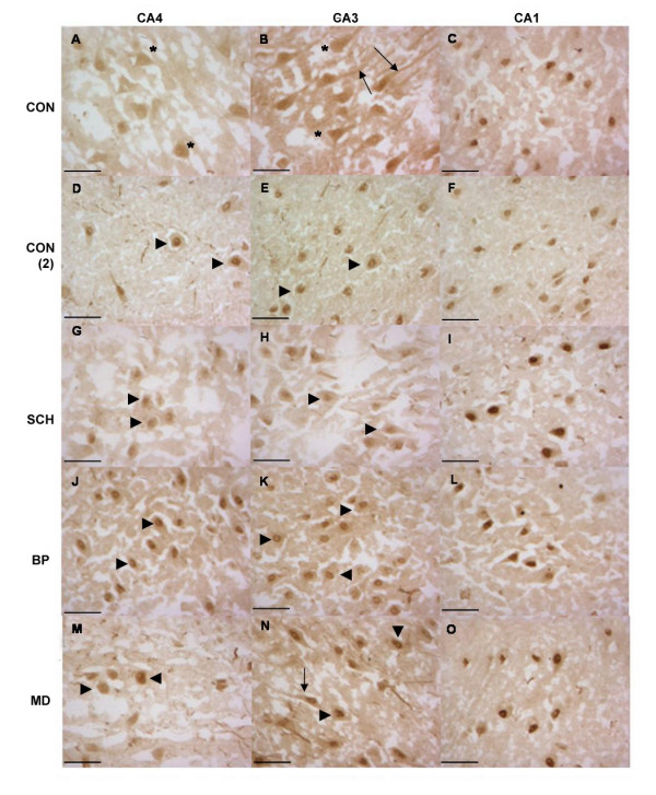
Figure 3.

Oct-6 Immunoreactivity in Fresh-frozen Tissue from Control, Schizophrenic, Bipolar Disorder and Major Depression. Oct-6 immunoreactivity was observed in all groups (CON A – C and CON (2) D- F), schizophrenics (SCH G – I), bipolar disorder (BP J – L) and major depression (MD M – O) and did not differ between groups or in comparison to controls. Oct-6-positive cells had the morphological appearance of pyramidal neurons. In the CA4 and CA3 regions immunoreactivity generally took on a nuclear localization (arrowheads) thought in some cases a more dispersed pattern of expression was noted (asterisks). Oct-6 immunoreactivity was also noted the apical dendrites of some cells (arrows). Though staining was robust throughout, it appeared stronger in the CA1 region (C, F, I, L & O), in all cases Oct-6 immunoreactivity in the CA1 was nuclear. Scale bars = 50 μm
