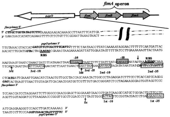
FIG. 2.
Schematic representation of the orientation of the pepO gene and the fimA operon in S. parasanguis. The expanded region shows the nucleotide sequence encompassing the 5′ coding region of pepO, the intergenic region between pepO and the fimA operon, and the 5′ coding region of the fimA operon. The fimA operon sequence (top sequence) and pepO sequence (bottom sequence) are shown. Sequence reported elsewhere (12) (dashed and dotted line) and sequences to which designated primers were designed for PCR and cloning purposes (bold italic type) are indicated. The arrows indicate the location of the start(s) of transcription for either the fimA operon or pepO gene, as determined by primer extension analysis. For the genes of the fimA operon and pepO gene, the start site of translation (bold type) and putative ribosomal binding sites (RBS) (underlined sequence) are shown. The putative −10 and −35 sites of the fimA operon (boxed sequence on grey background) and the pepO promoter (boxed sequence on white background) are indicated.

