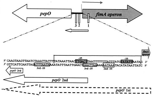
FIG. 3.
Schematic diagram of the S. parasanguis pepO gene and the fimA operon. The start of transcription of the fimA operon (thick black line) and the three start sites of pepO transcription (thin white boxes) are indicated. The expanded region shows the area of overlap between the fimA operon promoter and the pepO promoter. The fimA operon sequence (top sequence) and pepO sequence (bottom sequence) in the designated 5′ to 3′ orientations are shown. The region that shares high DNA sequence identity with the metallorepressor binding domain in S. gordonii sca operon (20) is boxed on three sides. The putative −10 and −35 sites of the fimA operon (boxed sequence on grey background) and the pepO promoter (boxed sequence on white background) are indicated. The arrows represent transcription products from either the fimA operon promoter or the pepO promoter.

