
Fig. 1. Nucleotide and amino acid sequences of mouse suprabasin.
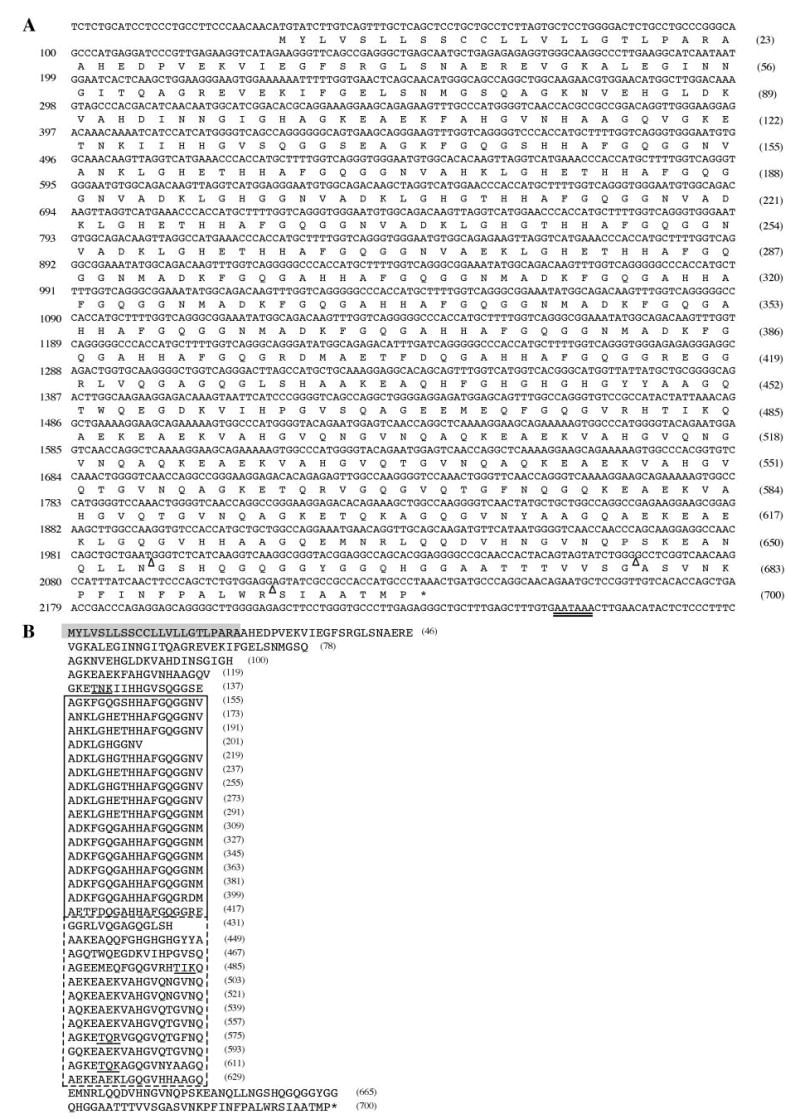
A, the nucleotide sequence of mouse suprabasin cDNA (2277 bp) was aligned with the predicted open reading frame amino acid sequence. Three open triangles indicate the sites of splicing (exon/intron boundaries), and the double-underlined nucleotides indicate the canonical poly(A) addition signal. B, the amino acid sequence of mouse suprabasin has been aligned to show the central repetitive domain region (boxed). The dotted line indicates the different repeat sequence region. The shaded residues highlight a predicted transmembrane region domain. The underlined sequences in the COOH-terminal domain indicate potential protein kinase C phosphorylation sites. Numbers in parentheses at the right side indicate number of residues.