
Fig. 4.
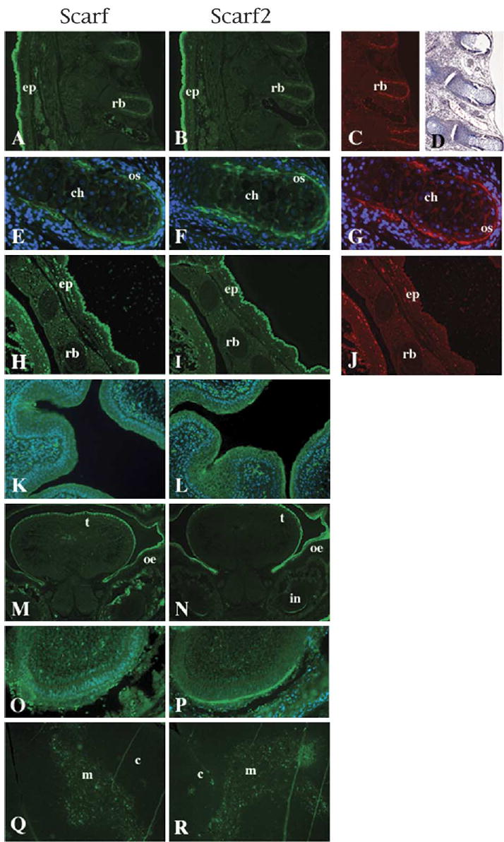
Scarf and Scarf2 protein expression. Dorsal view of sagittal sections of 16-day embryos treated with anti-Scarf (A), anti-Scarf2 (B), anti-OC (C) and corresponding Trichrome (Masson) staining (D). Treatments for OC and Trichrome staining were performed on serial sections. (E–G) 20× magnification of the dorsal aspect of rib bone, anti-Scarf (E), anti-Scarf2 (F) and anti-OC (G), with DAPI used for nuclear counterstain. The ventral aspects of sagittal section of corresponding (A)–(C): anti-Scarf (H), anti-Scarf2 (I) and anti-OC (J). (K–L) Scarf and Scarf2 staining in the epithelial cells of the bladder counterstained with DAPI. (M–N) Cross-sections of the head of 16-day embryo stained with anti-Scarf (M) and anti-Scarf2 (N). (O–P) 20× magnification of the incisor region from panels M and N, respectively. (Q–R) Adult thymus sections were stained with anti-Scarf (Q) and anti-Scarf2 (R), showing detection in the medullar region of thymus. Ep, epidermis; rb, rib bone; ch, chrondrocyte; os, osteoblast; t, tongue; oe, oral epithelia; in, incisor tooth; c, cortex; m, medulla.