
Figure 1. Stem cells in the interfollicular stratified epidermis and the bulge region of the hair follicle.
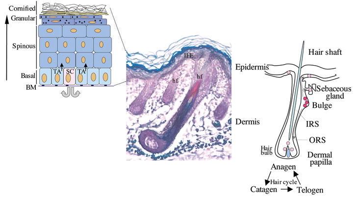
Left-hand panel, schematic representation of the stratified layers of the epidermis (basal, spinous, granular and cornified layers). The proliferative basal layer is adjacent to the basement membrane (BM). Stem cells (SC) generate transit amplifying (TA) cells which will differentiate to form the stratified layers. Middle panel, section of mouse skin stained to distinguish the different compartments of the hair follicle (hf) (Magnification 10×). Right-hand panel, schematic representation of a hair follicle and hair cycle, with the multipotent SCs (red) localizing to the bulge region. These cells migrate to populate the bulb region of the follicle, the sebaceous gland and the interfollicular epidermis (IFE; pink). IRS, inner root sheath; ORS, outer root sheath.