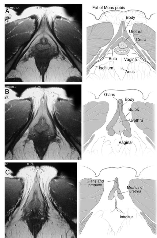
Fig. 1.

A, clitoris and its components, including bulbs, crura and corpora, are well demonstrated in axial plane. These structures lie ventral and lateral to urethra and vagina as cluster or complex. MRI specifications for this scan were FSE, TR:4000, TE:15/Ef, EC:1/1 16kHz, FOV: 16x16, 4.0thk/1.0sp, 30/04:16, 256x256/2 NEX, FCs/NP. B and C, next 2 sections caudal to section A in same volunteer. B, clitoral glans ventral to remainder of clitoris. Its midline septum and prepuce are evident. C, most caudal section reveals glans and caudal limit of urethra (urethral meatus), clitoral bulbs and vagina (introitus). In this perineal section clitoral body and crura are not present and urethral meatus and vaginal introitus are not distinct. MRI specifications were FSE, TR:4000, TE:15/Ef, EC:1/1 16kHz, FOV:16x16, 4.0thk/1.0sp, 30/04:16, 256x256/2 NEX, FCs/NP.
