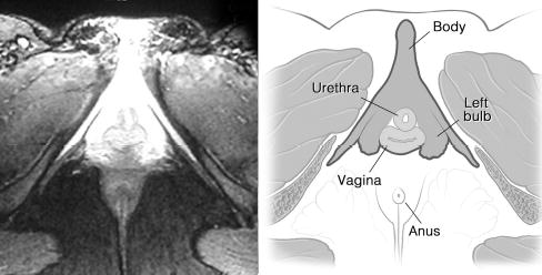
Fig. 2.

Using fat saturation highlighted cavernous tissue of clitoris surrounding urethra and vagina, while other structures appeared gray or black. Triangular clitorourethrovaginal complex was clearly seen using this sequence. MRI specifications were FSEIR, TR:4083, TE:22/Ef, EC:1/1 31.2kHz, TI:165, FOV20x20, 6.0thk/1.5sp, 15/06: 32, 256x192/4 NEX, NP/VB/SQ/SPF.
