
Figure 4. Lung metastasis signature in human primary breast tumours.
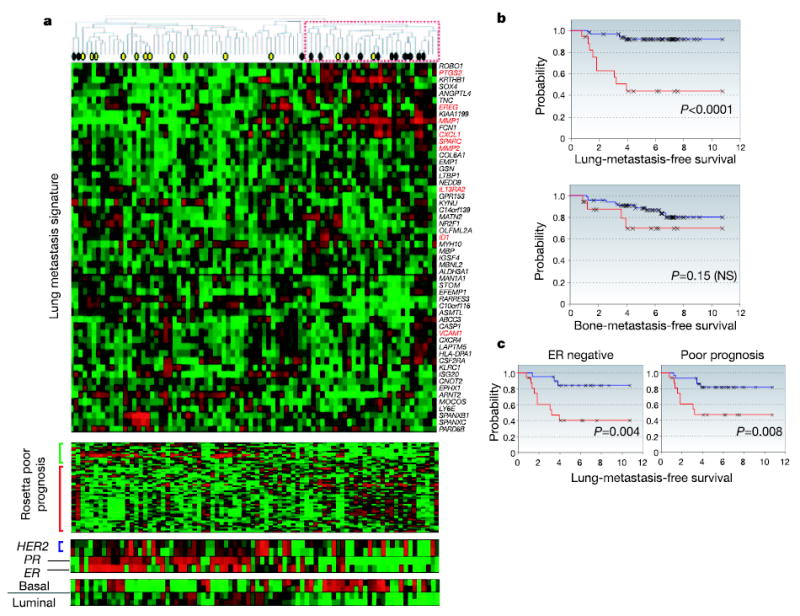
a, Hierarchical clustering of primary breast carcinomas from a cohort of 82 breast cancer patients was performed with the 54 lung metastasis signature genes. A dendrogram of the tumours is shown at the top, with tumours from patients who developed lung metastasis (black circles) or metastasis at non-pulmonary sites (yellow circles) denoted. A subcluster with a reproducibility index of 0.71 (dashed red box) groups tumours that tended to express the lung metastasis signature in a manner resembling the LM2 cell lines. The genes were also clustered; gene names are on the right. Functionally validated genes are in red. The Rosetta poor-prognosis signature is displayed with the genes underexpressed (green bar) and overexpressed (red bar) in poor-prognosis tumours indicated at the left. The expression of HER2, progesterone receptor (PR), oestrogen receptor (ER) and basal and luminal keratins is shown. Expression of the lung metastasis signature was confirmed in the independent Rosetta breast cancer cohort (Supplementary Fig. S6). b, Lung-metastasis-free survival and bone-metastasis-free survival for MSKCC patients who either expressed (red line) or did not express (blue line) the lung metastasis signature based on a classifier trained with the Rosetta cohort (Supplementary Fig. S7 and Supplementary Methods). The P value for each survival curve is shown. c, Lung-metastasis-free survival restricted to patients with ER-negative tumours or Rosetta-type poor-prognosis tumours.