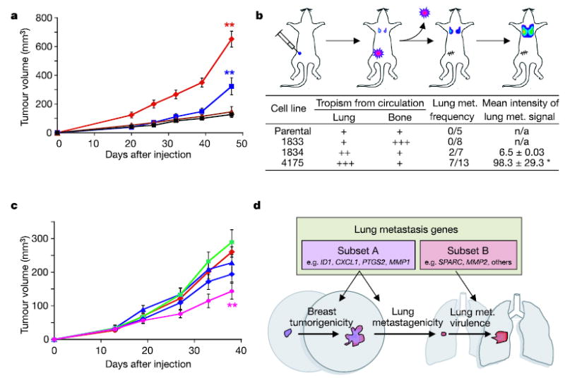
Figure 5. Breast tumorigenicity and lung metastagenicity partially overlap.

a, Representative MDA-MB-231 cell populations were injected into the mammary fat pad of immunodeficient mice and monitored for tumour growth. Red diamonds, 4175 cells (n = 9, where n is the number of mice in each cohort); blue squares, 1834 cells (n = 10); brown triangles, 1833 cells (n = 5); black squares, parental cells (n = 5). Each curve shows tumour volumes in cubic millimetres (means ± s.e.m.). b, As shown in the diagram, mice were inoculated with the indicated MDA-MB-231 cells into the mammary fat pad and tumours were removed after reaching a volume of 300 mm3. Lung metastasis was monitored with BLI, and normalized photon flux was measured 2 weeks after removal of the primary tumour. Asterisk, a mouse in the 4175 cohort with an unusually high normalized photon flux of 36,400 was excluded. c, Growth in mammary fat pad of highly lung- metastatic 4175 (LM2) cells after stable shRNA knockdown of the following gene products: red diamonds, shControl; blue triangles, shVCAM; green squares, shIL13RA2; blue diamonds, shSPARC; pink circles, shID1. shControl refers to a cell line transduced with a short hairpin construct that did not result in effective knockdown of its target gene. Two asterisks, P < 0.01 by a one-sided rank test. Each curve shows tumour volumes in cubic millimetres (means ± s.e.m.). d, A model of two classes of genes contained within the lung metastasis signature. The first class (subset A) confers both breast tumorigenicity and basal lung metastagenicity. Examples may include ID1, CXCL1, PTGS2 and MMP1. The second class (subset B) confers functions specific to the lung microenvironment, facilitating lung metastatic virulence. Examples may include SPARC and MMP2.
