
Figure 3.
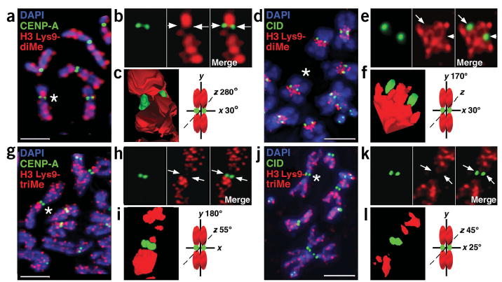
H3 is not di- or trimethylated at Lys9 in CEN chromatin at metaphase. (a–j) Immunofluorescence patterns on human (a,g) and D. melanogaster (d,j) metaphase chromosomes show localization of H3 Lys9-diMe or H3 Lys9-triMe (red) relative to CENP-A and CID staining (green). Chromosomal DNA was stained with DAPI (blue). (b,e,h,k) Enlarged views of chromosomes marked with asterisks in a, d, g (human chromosome 1) and j (D. melanogaster chromosome 3), with merged images to the right of the single-color images. Arrows point to the areas on individual chromosomes where CENP-A/CID is located. (c,f,i,l) Three-dimensional modeling of antibody staining on the same human and D. melanogaster metaphase chromosomes from b, e, h, and k. Chromosome cartoons depict the degree to which each three-dimensional model was rotated around the x-, y- and/or z-axes. In all cases, CENP-A/CID shows minimal overlap with H3 Lys9-diMe or tri-Me staining. H3 Lys9-diMe was present in the spaces between the CENP-A/CID cylinders15 on the two sister chromatids, as well as on either side of the centromere region along the chromosome arms. In contrast, H3 Lys9-triMe was offset significantly from CENP-A/CID staining along the chromosome arms, and was not present in the spaces between the sister centromeres. Quantifications of overlaps are shown in Figure 4. Scale bars, 15 μm in a,g; 5 μm in b,j.