
Figure 6.
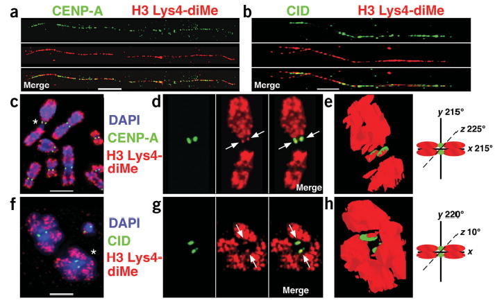
H3 is dimethylated at Lys4 in CEN chromatin. (a,b) Extended chromatin fibers from human cells (a) and D. melanogaster S2 cells (b) were stained with antibodies to CENP-A/CID and H3 Lys4-diMe, detected with secondary antibodies conjugated with FITC (green) and Cy3 (red), respectively. Single-color images are shown separated from the merged image. H3 nucleosomes interspersed with CENP-A/CID nucleosomes are dimethylated at Lys4 in both humans and flies. (c–h) Immunofluorescence patterns on human (c) and D. melanogaster (f) metaphases stained with H3 Lys4-diMe antibodies (red). DNA was stained with DAPI (blue). Antibody localization on metaphase chromosomes shows that H3 Lys4-diMe is located to close to, and partially overlapping with, CENP-A/CID (green). (d,g) Enlarged view of chromosomes marked with asterisks in c and f, with single-color images to the right of the merged images. (e,h) Three-dimensional modeling of antibody staining on individual metaphase chromosomes from d and g shows that H3 Lys4-diMe is located in the region between sister kinetochores, partially overlapping with CENP-A/CID staining (arrows), consistent with the coiling or looping model proposed for CEN chromatin higher ordering packaging at metaphase15. The asterisk in d denotes human chromosome 1, which is particularly under-represented for H3 Lys4-diMe in the 1q region. Chromosome cartoons depict the degree to which each three-dimensional model was rotated around the x-, y- and/or z-axes in e and h. Fiber and metaphase results are quantified in Figures 2 and 4. Bars, 15 μm in a,c; 5 μm in b,f.